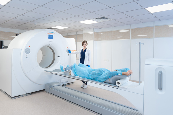
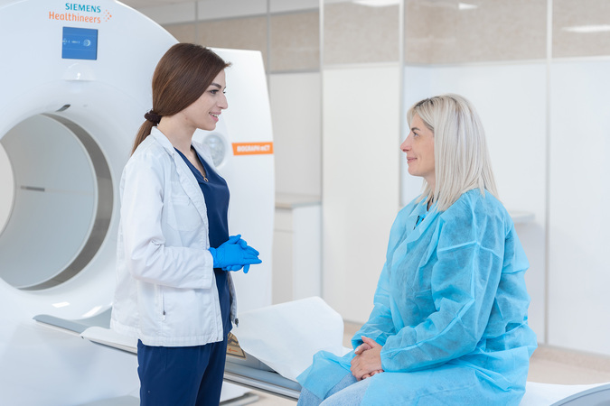
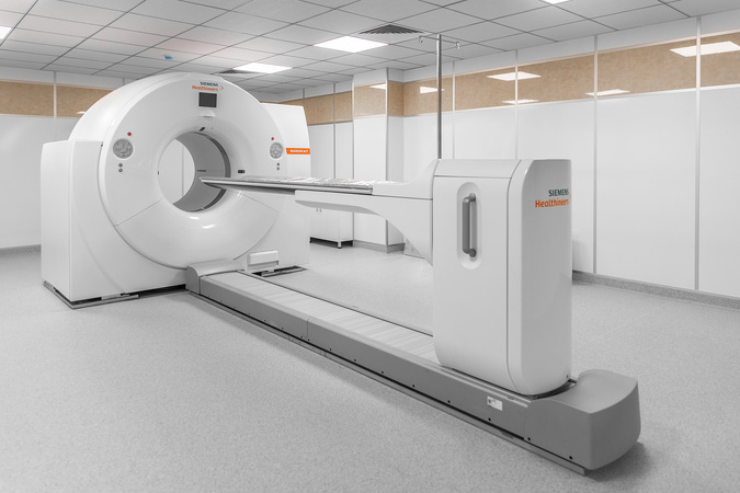
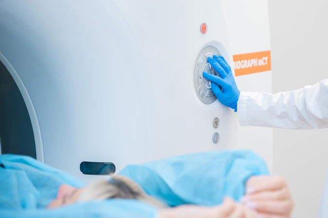
The latest generation Biograph mCT PET/CT machine by Siemens at the "Erebuni" Radiotherapy Center

Positron emission tomography (PET) is one of the most important modern diagnostic methods in the field of oncology. It is performed using radioisotopes, which require special facilities and stringent safety measures. At the “Erebuni” Radiotherapy Center, where this advanced diagnostic method has recently been implemented, all conditions have been created for conducting examinations safely and effectively.
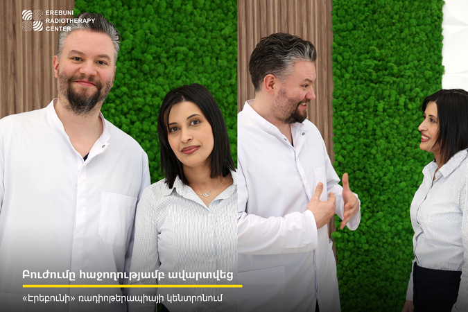
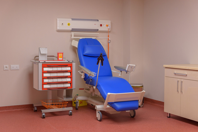
“We have a laboratory where radioactive materials are prepared for injection into patients. A doctor prescribes an individual dose for each patient, and the lab technician prepares the radioactive material for injection according to that dose. After the injection, the patient spends about an hour in the examination room to allow the radioactive material to accumulate in the body,” says Gurgen Elbakyan, head of the radionuclide diagnostics department at the “Erebuni” Radiotherapy Center.
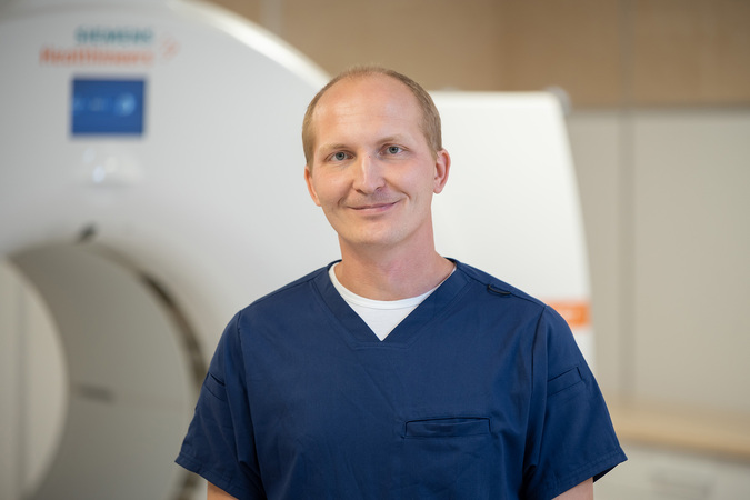
“During this entire time, the patient should not move, talk, or read. In other words, it is essential to completely avoid muscle tension. This is because the radioactive material can accumulate not only in tumor tissues but also in muscles, which can interfere with our image interpretation. Half an hour before the examination starts, the patient is advised to drink about a liter of water to ensure proper distribution of the radioactive material,” adds Pavel Gelezhen, a radiologist at the “Erebuni” Radiotherapy Center.
The radioisotope administered to the patient resembles sugar. It has the ability to bind to tumor cells, allowing the physician to see the accumulation of these cells in the body. Before obtaining the isotope, it is necessary to check the patient’s blood glucose level, as a high sugar content can distort the image.
“In 90, and even 95% of cases, we use positron emission tomography to stage oncological processes.
This means that when a patient unfortunately discovers they have a malignant tumor, we need to determine how to treat it, and for that, we need to find out if the patient has metastases and how many there are. Rarely, but sometimes we use positron emission tomography to locate the primary tumor in cases where metastatic tumors are found in the patient’s body but the origin or primary site is unknown,” says Gelezhen.
Positron emission tomography is also used to evaluate the effectiveness of cancer treatment, which is especially important in the case of lymphomas. Previously, assessing treatment response required surgical biopsy of lymph nodes. Now, PET allows us to avoid that.
After spending an hour in the examination room, the patient is placed into the PET/CT scanner. The examination is conducted in two phases: first, a computed tomography (CT) scan is performed, followed by the positron emission tomography.
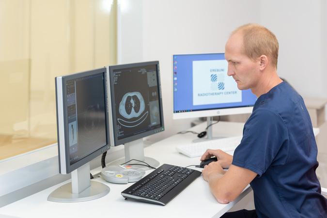
“Initially, we conduct a CT scan to see the anatomy of the person, the bones, soft tissues, and organs. The next step involves collecting information on the distribution of the radioactive material. The third, crucial phase is the fusion of the two images, allowing us to clearly see where tumors are located and where they are not,” explains the radiologist.
How safe is this method of investigation, considering that a radioactive substance is injected into the patient? Specialists explain that during PET examinations, a radioisotope with a very short half-life is used.
“The advantage of the radioactive material we use here in the clinic is its short half – partition phase, which is the time it takes for the substance to decay and turn back into regular glucose.
The short half – partition phase is about two hours, meaning that within two hours, half of the injected material completely disappears,” says the doctor.
Practically, by the end of the examination, only a very small amount of this substance remains in the patient’s body. However, for added safety, the patient remains in a post-examination room for about an hour.
The center's building is constructed according to all safety standards for nuclear medicine. The rooms are lead-lined, and the drainage and ventilation systems are designed with radiation regulations in mind.
At the “Erebuni” Radiotherapy Center, the latest generation Siemens Biograph mCT PET/CT 64 device is used. It has several advantages, the key one being a continuously moving table.
“Typically, positron emission tomography scanners use a sequential movement of the table. As a result, to obtain optimal image quality, more radioactive material must be injected. Our device utilizes a continuous motion feature, allowing us to inject less radioactive material, reducing the patient’s exposure to radiation, while simultaneously improving image quality compared to previous generation devices,” says Gelezhen.
The launch of this new generation positron emission tomography device in Armenia is of tremendous importance. The “Erebuni” Radiotherapy Center is equipped with all modern methods of radiation treatment for cancer, and its specialists have undergone international training, with many invited from foreign countries. The center is ready to provide high-quality radiation therapy to both Armenian citizens and foreign nationals.
Other news
Our specialists continuously advance their knowledge.
Read more
The process of radiation therapy begins with the right questions and clear answers
Read more
Media Visit to the “Erebuni” Radiotherapy Center
Read more
Sergey Golub answered frequently asked questions
Read more
Interview with the Head of the Erebuni Radiation Therapy Center
Read more
Interview with the Head of the Radiation Therapy Department at the Erebuni Center
Read more