Computed tomography with topometry
This service includes only CT simulation without further work from radiation therapists and medical physicists. This may be necessary if the patient plans to undergo treatment at another clinic.
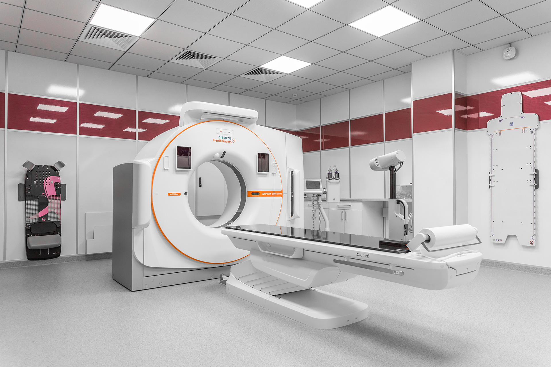
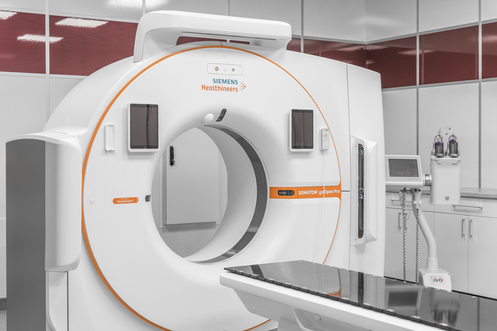
One of the key stages in preparing for radiation therapy is CT marking (CT topography), which involves performing a computed tomography scan. This preparatory step is necessary to obtain detailed images of internal organs and tissues, which are needed for precise planning of the radiation exposure and controlling the radiation doses affecting healthy tissues.
At the beginning of the CT marking, the patient is familiarized with the process. Specialists explain how the procedure will go, why it is necessary, and what sensations the patient might experience. Then, special immobilization devices are selected to ensure the patient remains still during radiation therapy sessions. The choice of device depends on the treatment area and individual patient characteristics. These may include vacuum mattresses, thermoplastic masks, and head fixation devices, among others. These devices help the patient remain in a fixed position in comfortable conditions, and this position will be replicated at each treatment session.
After the fixation devices are prepared, a scan is conducted using the CT scanner according to the topography protocol, and centering marks are applied to the devices or the patient's body. After scanning, photos of the patient may also be taken to document the positioning details. An important aspect of the preparation is creating a comfortable and trusting environment, as well as providing emotional support.
Options for CT Marking:
- CT marking with contrast: This method is used to improve image detailing, especially when the tumor is near important blood vessels or organs. The contrast agent helps to clearly visualize anatomical structures and, in some cases, allows for more accurate determination of the radiation volume.
- CT marking without contrast: This option is applied when additional visualization is not required due to tumor localization or if the patient has contraindications for contrast material. Despite the lack of contrast enhancement, this method still allows for the creation of high-quality radiation therapy plans.

Consultation with a Radiation Therapist
After CT marking, the patient consults with a radiation therapist, who is responsible for the following preparation stages:
- Determining the treatment method: The doctor selects the appropriate type of radiation therapy depending on the type and stage of the disease. This may involve external beam radiation therapy or brachytherapy (internal radiation therapy).
- Developing the treatment plan: Based on the CT marking data, an individualized treatment plan is created, which specifies the number of treatment sessions and the dose of radiation delivered during each session.
- Determining the radiation volume: The specialist outlines the treatment zones to minimize the impact on healthy tissues.
- Dosimetric planning: In collaboration with medical physicists, dosimetric planning is carried out, and a personalized radiation therapy plan is created for the patient, ensuring maximum therapeutic effect with minimal side effects.
Preliminary preparation for radiation therapy is a comprehensive and crucial stage in treatment, involving the collection of medical documents, performing CT marking, and consulting with a radiation therapist. These measures help create a personalized treatment plan, ensuring high precision, safety, and comfort for the patient.

