

Positron Emission and Computed Tomography PET/CT Ga-68 PSMA
Ga-68 PSMA PET/CT is a high-precision examination used to detect tumor lesions that express PSMA, assess the extent of disease, and support treatment planning. The method is especially widely used in the diagnosis and follow-up of prostate cancer, as PSMA is highly expressed on the surface of prostate cancer cells.
ㅤ
ㅤ
ㅤ
In modern oncological diagnostics, it is important not only to see structural changes in organs, but also to understand the biological activity of suspicious lesions. This is exactly what PET/CT makes possible by combining the molecular information of positron emission tomography with the anatomical imaging of computed tomography.
What is Ga-68 PSMA PET/CT?
Ga-68 PSMA PET/CT is an imaging examination that uses a special radiopharmaceutical labeled with the Gallium-68 radionuclide. This substance binds to a protein called PSMA. PSMA may be highly expressed on the surface of certain tumor cells, making them visible on PET/CT imaging.
The PET component shows active areas where the radiopharmaceutical accumulates, while CT helps determine their exact anatomical location. As a result, the physician receives not just an image, but important information about the possible extent, activity, and location of the disease.
How the method works
During the examination, the patient receives an intravenous injection of the Ga-68 PSMA radiopharmaceutical. It circulates through the bloodstream and binds to cells that have high PSMA expression on their surface. After a certain waiting period, the scan is performed.
One of the key advantages of PET/CT is that it provides two types of information at the same time: functional and anatomical. This is especially important when the correct treatment strategy depends on an accurate assessment of how far the disease has spread.
How Ga-68 PSMA PET/CT differs from CT and MRI
Computed tomography and magnetic resonance imaging are important and widely used diagnostic methods. They help assess the structure of organs, their size, tissue changes, enlarged lymph nodes, and other anatomical features. However, in some cases, small tumor lesions or early-stage changes may still be difficult to detect using structural imaging alone.
Ga-68 PSMA PET/CT evaluates not only structure, but also activity at the cellular level. For this reason, it may be more sensitive in situations where the disease is still small in volume but already shows uptake of the PSMA radiopharmaceutical.
Thanks to these differences, Ga-68 PSMA PET/CT is considered an important modern method of molecular imaging, which can complement or clarify the results of CT, MRI, and other examinations.
Main advantages of Ga-68 PSMA PET/CT
One of the main advantages of Ga-68 PSMA PET/CT is its high sensitivity. The examination can help detect small lesions that may still be difficult to see with conventional imaging methods.
Another advantage is the possibility of whole-body assessment. A single examination can provide information about different anatomical areas, depending on the purpose of the scan and the medical indications.
A third important advantage is its impact on treatment planning. Accurate diagnosis can help the physician choose a more targeted approach, such as local treatment, radiation therapy, systemic treatment, surgery, or a combined strategy.
The examination is also valuable during follow-up after treatment. If laboratory or clinical data suggest possible disease activity, Ga-68 PSMA PET/CT can help clarify whether there is a local lesion, lymph node involvement, or disease spread.
Who may need this examination and when?
Ga-68 PSMA PET/CT is performed based on a physician’s referral. The need for the examination is determined individually, taking into account the patient’s diagnosis, previous imaging results, laboratory data, and treatment history.
The examination may be indicated when it is necessary to clarify the extent of a tumor disease, evaluate the nature of suspicious lesions, identify the cause of changes after treatment, or plan further treatment.
In prostate cancer, Ga-68 PSMA PET/CT is most commonly used for disease staging, detection of possible recurrence after treatment, clarification of the cause of rising PSA levels, and detection of metastatic lesions.
How to prepare for Ga-68 PSMA PET/CT
Patients are usually advised to drink enough water before the examination. It is also necessary to avoid food intake for 4 hours before the scheduled examination time.
On the day of the examination, it is recommended to wear comfortable clothing and, if possible, avoid metal items such as jewelry, belts, clothes with metal buttons, and similar accessories.
How the examination is performed
The examination begins with an initial assessment. The specialist reviews the patient’s medical documents, previous examination results, laboratory data, and treatment history. This is important for the correct interpretation of the scan results later on.
The patient then receives an intravenous injection of the Ga-68 PSMA radiopharmaceutical. After the injection, the patient needs to wait for some time so that the substance can circulate through the body and accumulate in the relevant areas. During this period, the patient remains in a calm and comfortable environment.
During the scan, the patient lies on the tomography table. It is important to remain as still as possible, as movement can affect image quality. The scan itself usually takes a relatively short time, but the entire process — including preparation, injection, waiting time, and scanning — may take several hours.
How the results are analyzed
The results of Ga-68 PSMA PET/CT are analyzed by a specialist. The physician evaluates the areas of radiopharmaceutical uptake, their location, uptake intensity, and correlation with clinical data.
The report may describe the location of suspicious lesions, their size, degree of uptake, and possible clinical significance. It is important to understand that PET/CT results should not be interpreted in isolation, but together with the patient’s full medical picture: previous examinations, laboratory values, diagnosis, and treatment history.
Safety and contraindications
Ga-68 PSMA PET/CT is considered a safe diagnostic examination when performed according to medical indications and appropriate protocols. A small amount of radiopharmaceutical is used during the procedure, and the radiation exposure is assessed so that the diagnostic benefit of the examination outweighs the potential risks.
The Ga-68 radionuclide has a short half-life, and the radiopharmaceutical is mainly eliminated from the body through urine. For this reason, after the examination, patients are advised to drink enough fluids and empty the bladder frequently.
Contraindications and limitations are assessed individually. They may be related to the patient’s general condition, inability to lie still for a prolonged period, impaired kidney function, or the need for contrast-enhanced CT.
Ga-68 PSMA PET/CT at Erebuni Radiotherapy Center
At Erebuni Radiotherapy Center, Ga-68 PSMA PET/CT is performed for high-precision molecular diagnostics. The center’s approach is based on modern equipment, professional expertise, and individual assessment of each patient’s case.
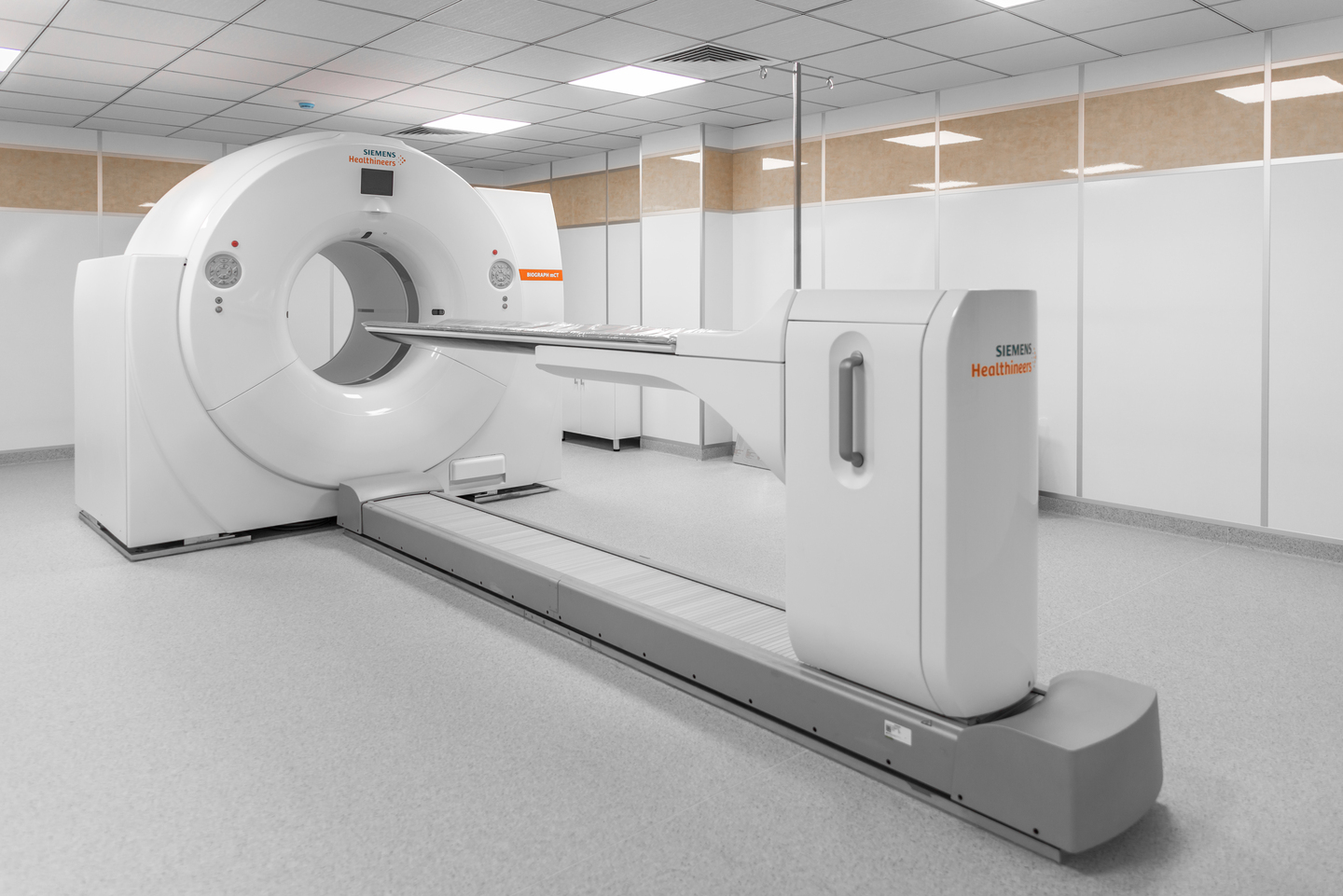
Erebuni Radiotherapy Center uses the modern Siemens Biograph mCT 64 PET scanner. The examination is performed in accordance with medical protocols, ensuring high image quality, patient safety, and professional interpretation of the results.
Where to undergo Ga-68 PSMA PET/CT in Yerevan and Armenia
Ga-68 PSMA PET/CT is available in Yerevan at Erebuni Radiotherapy Center. The center serves patients from Armenia and abroad, providing modern diagnostics, a professional approach, and individual support throughout the entire examination process.